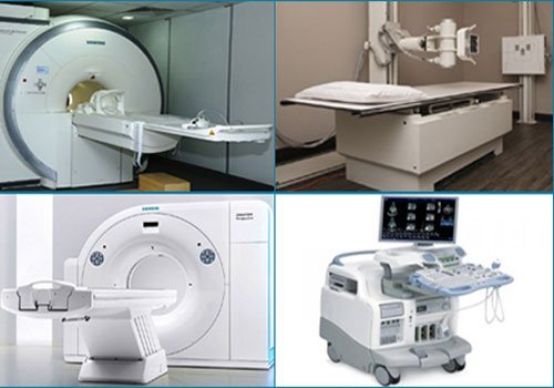
Radiology is the branch of medicine that uses imaging techniques to diagnose and treat diseases that cannot be seen from outside the body. A variety of imaging techniques, such as X-ray Radiography, Ultrasound, Computed Tomography (CT), Nuclear Medicine and Magnetic Resonance Imaging (MRI) are employed for diagnosing and/or treating various complicated diseases. Interventional Radiology follows medical procedures in line with standard procedures of Imaging Technologies.
Radiographers are often also known as Diagnostic Radiologists. They are responsible for interpreting or ‘reading’ medical images and producing brief and concise reports based on them. They analyze the findings and impressions of such diagnoses. Such reports are then referenced to the Clinician, either routinely or on emergency-basis. The results thus obtained of the Imaging procedures are stored digitally in PACS (Picture Archiving and Communication System).
Radiological examinations are rarely indicated in the acute phase of a urinary tract infection. An exception is when an obstruction of a ureter is suspected in a patient with signs of pyelonephritis. In children with pyelonephritis or with recurrent cystitis, radiological examinations for identification of congenital anatomical defects and/or ureteral reflux should be performed after treatment of the acute infection. For detection of vesicoureteral reflux in children, contrast-enhanced ultrasound is recommended as a better alternative to micturating cystourethrography.
In adults who have recovered from pyelonephritis it is recommended that ultrasound or a radiological examination is performed to exclude renal scars from childhood episodes of pyelonephritis.